Figure 4.
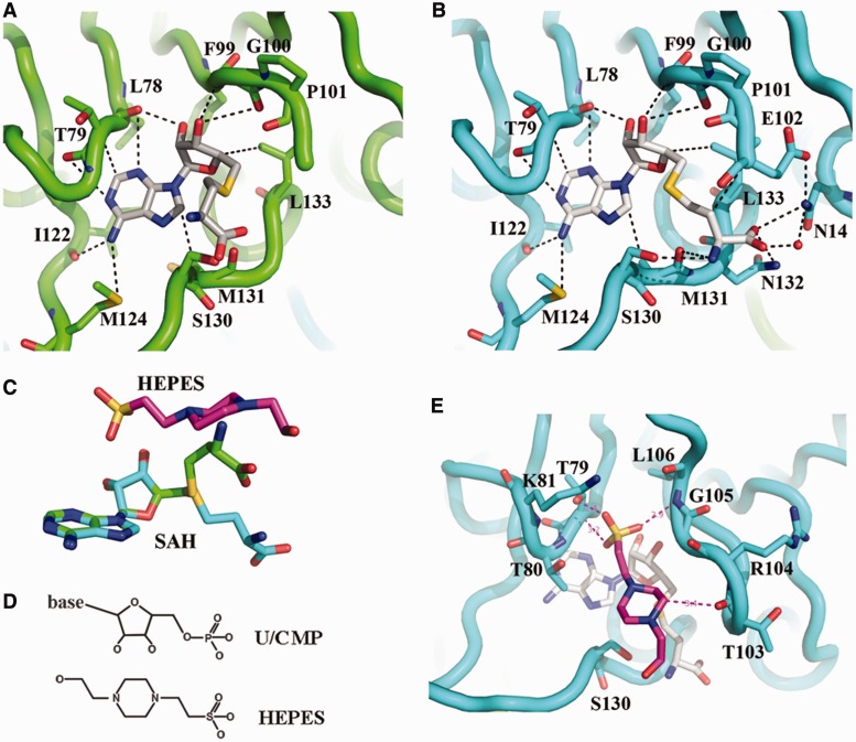
SAH and HEPES binding. (A) SAH bound in subunit A. The carbon atom of SAH is shown in white, the backbone of EcTrmL is in green, and all the residues within 4 Å from SAH are shown in stick. (B) SAH binding details in subunit B, the backbone of EcTrmL is shown in cyan. (C) The crystal structures of SAH molecules from subunit A (green) and subunit B (cyan) are superimposed and shown as sticks, with the structure of HEPES in magenta. (D) The chemical structure of the ribose and phosphate of U/CMP, and the HEPES molecule. (E) The structure of a HEPES molecule bound to EcTrmL, with all the residues within 4 Å shown as sticks. The carbon atoms of HEPES and SAH are shown in magenta and white, respectively.

