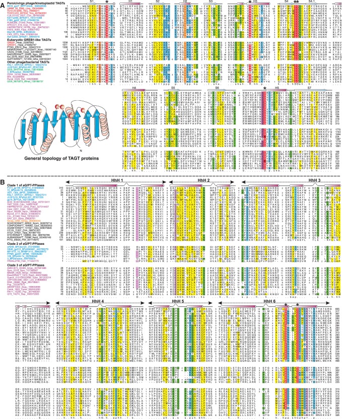
Figure 3.
Multiple sequence alignment of the DNA base glycosyltransferases (A) and the aG/P-T-pyrophosphorylases (B). Protein sequences are labeled by gene names followed by species abbreviation and Genbank GIs. Phage protein names are colored blue, bacterial ones in pink, archaea in orange and eukaryotes in black. Predicted catalytic residues for both TAGT and aG/PT-PPlase are indicated by asterisks, with secondary structure assignments shown above the alignment. Alignment columns are colored based on the 80% conservation consensus. Topology of the glycosyltransferase domain is adjacent to the alignment. See Supplementary Data for species abbreviations.

