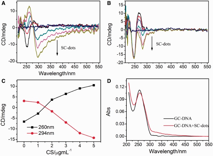
Figure 2.
CD spectra of 2 µM GC-DNA (A) and AT-DNA (B) titrated with different SC-dots: 0, 1, 2, 3, 4, 5 µg/ml. The straight line in middle is CD spectrum of 5 µg/ml SC-dots alone. (C) Plot of CD intensity at the 260 nm (black square) or at 294 nm (red circles) as a function of the SC-dots concentration. The data were adopted from (A). (D) UV spectra of GC-DNA in the absence or presence of 2µg/ml SC-dots.

