
Figure 1.
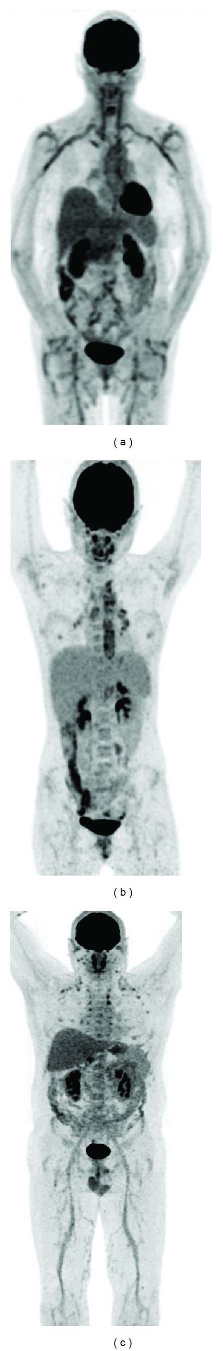
FDG-PET examples of vasculitis: (a) giant cell arteritis and polymyalgia rheumatica: high FDG uptake in the large vessels (aorta, subclavian arteries, carotid arteries, iliac arteries, and femoral arteries) accompanied by high uptake in the large joints (shoulders and hips), (b) Takayasu's arteritis: high FDG uptake located more centrally (aorta and main branches in the thoracic region) and in this case uptake in reactive lymph nodes in mediastinum and hili (confirmed by biopsy), and (c) polyarteritis nodosa and polychondritis: high uptake in the medium- and small-sized arteries (best visible in the legs) accompanied by uptake in the nose, the ears, and the costochondral regions.