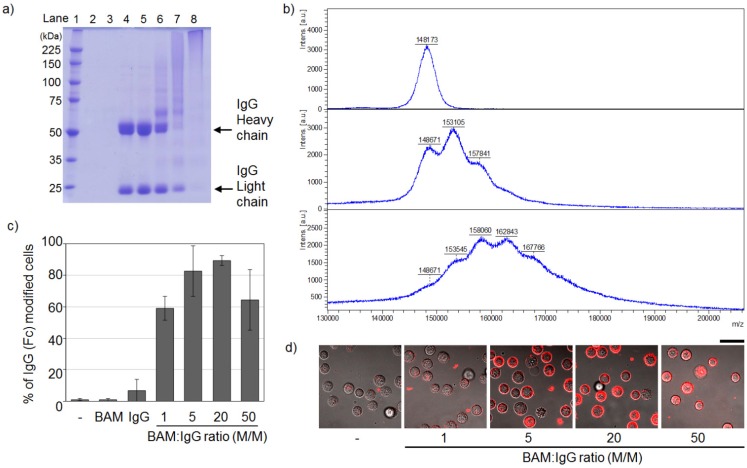
Figure 2.
Optimization of the condition of IgG-BAM conjugation by changing the final molar ratio of BAM to IgG. (a) Qualitative analysis of the attachment of BAM to IgG by a reducing 7.5% polyacrylamide SDS-PAGE gel stained with Coomassie brilliant blue. Lane 1, protein molecular weight markers in kDa; lane 2, PBS as a control; lane 3, BAM as a control; lane 4, IgG as a control; lanes 5−8; IgG treated with BAM at various ratios: 1:1, 1:5, 1:20 and 1:50; (b) MALDI-TOF MS spectra of intact IgG (top), IgG treated with BAM at the IgG:BAM ratio of 1:5 (middle) and 1:20 (bottom); (c) Qualitative analysis of the incorporation of IgG-BAM conjugates into the membrane of HeLa cells by flow cytometry. (n = 3); (d) CLSM observation. Bar = 40 mm.

