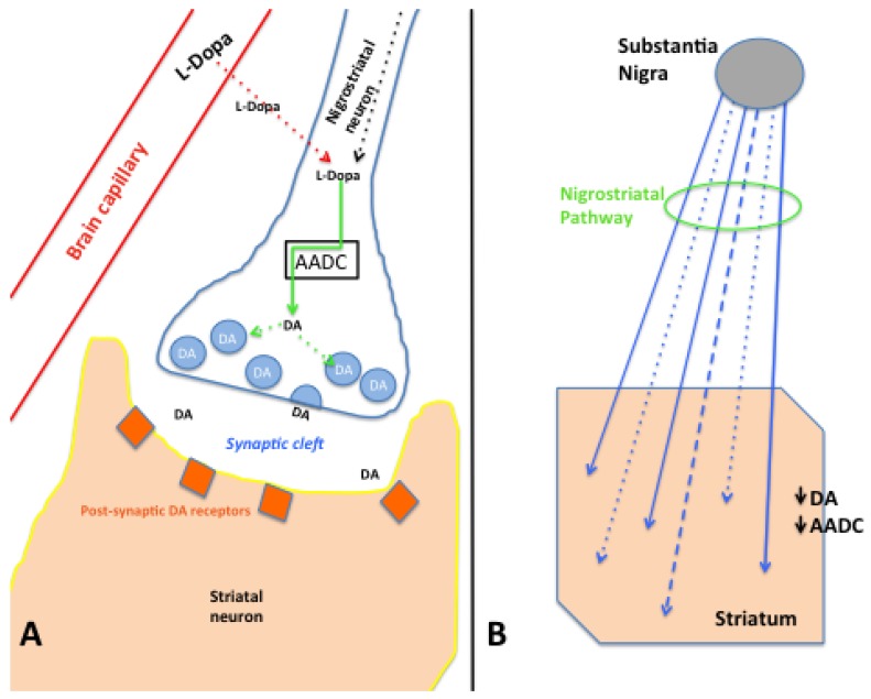
Figure 1.
(A) Schematic representation of a brain dopaminergic perisynaptic region. A local capillary allows blood supply to deliver L-dopa to the brain parenchyma, traversing the blood-brain barrier and being taken up by the presynaptic dopaminergic nigrostriatal neuron (dotted red arrow). These neurons also produce L-dopa within the cell and transport it to the presynaptic terminals (dotted black arrow). At the neuron terminal, AADC (black box) converts L-dopa to dopamine (DA) (solid green arrow), allowing DA to be packaged (dotted green arrows) into presynaptic vesicles (light blue circles) for release into the synaptic cleft as part of neurotransmission. Released DA is able to interact with postsynaptic receptors on striatal neurons; (B) Schematic of the degeneration of the nigrostriatal pathway. Nigrostriatal dopaminergic neurons (blue arrows) originate primarily within the midbrain substantia nigra (dark gray oval) and extend into the striatum. With progressive degeneration of the nigrostriatal pathway (green circle), fibers lose their normal caliber and eventually lose function (dashed blue line) and degenerate (dotted blue lines), in association with loss of dopaminergic perikarya in the substantia nigra. Striatal biochemical assays show decreased DA and AADC levels associated with nigrostriatal degeneration.

