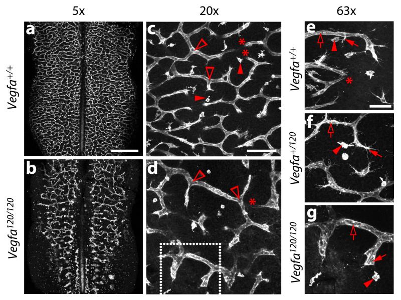
Fig. 5. Examples of vascular defects in the hindbrain of mice from Vegfa+/120 intercrosses.
E11.5 littermate hindbrains from timed matings of Vegfa+/120 parents were fluorescently labelled with IB4 and imaged at the indicated magnifications. Examples of vascular intersections (clear arrowheads) for branch point quantification and tissue macrophages (solid arrowheads) are indicated in (c,d). Examples of tissue macrophages (solid arrowheads), endothelial tip cells (solid arrows) and endothelial stalks cells (clear arrows) are indicated in (e-g). Areas indicated with asterisks show vessels that dive into deeper layers of the hindbrain and appear ‘cut-off’, because the confocal z-stack does not span the entire SVP. The area shown in (g) is a higher magnification of the area indicated with a white box in (d). Scale bars: (a,b) 500 μm; (c,d) 100 μm, (e-g) 50 μm. All animal procedures were performed in accordance with institutional and UK Home Office guidelines.

