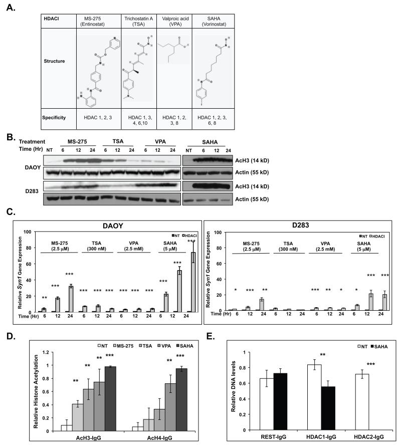
Figure 3. HDACIs upregulate Syn1 gene expression.
(A) Structure and specificities of HDACIs used in this study (30) (B) Western blot analysis and (C) Q-RT-PCR were performed to measure acetylation of histone H3 (AcH3) and changes in Syn1 gene expression respectively in DAOY and D283 in response to MS-275 (2.5 μM), TSA (300 nM), VPA (2.5 mM), or SAHA (5 μM) treatment for the indicated time-periods. Actin was used as a loading control for Western blots. Syn1 expression is reported relative to 18sRNA levels and compared with untreated controls (NT) set to 1. ChIP analyses were performed to compare changes in (D) acetylation of histones H3 and H4 or (E) REST, HDAC1 and HDAC2 binding to the Syn1 RE1 element in DAOY cells following HDACI treatment. Samples were normalized to input DNA and compared with DNA pulled down by non-immune sera (IgG). Assays were performed in triplicate and results reported as mean +/− standard error. Statistical significance is shown as *(0.1 >p>0.05), **(0.05≥p>0.01) or ***(p≤0.01).

