
Figure 2. Effect of weekly pulse dosing rapamycin on autophagy and a1AT protein in the liver in PiZ mice.

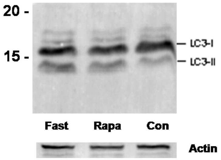
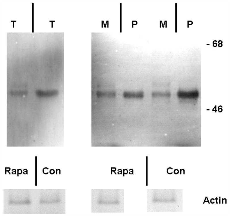
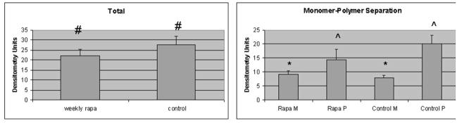
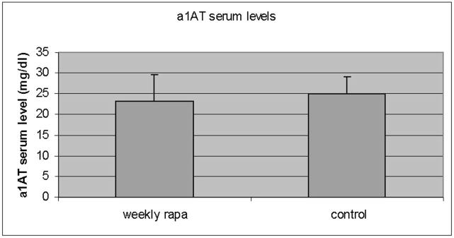
Panel a, percent area of hepatocellular cytoplasm occupied by autophagic vacuoles in PiZ mice treated with weekly dosing rapamycin versus vehicle control PiZ mice (p<0.5 by ANOVA). Bars +/− S.D. Panel b, representative example of SDS-PAGE followed by immunoblot for LC3 from a WT mouse liver fasted 12 hours (fast) as a positive control, PiZ mouse liver treated with weekly dose rapamycin (rapa) or vehicle control (con) of total hepatic protein lysate. LC3-I and LC3-II reactive bands are shown. Actin loading controls as shown, below, and molecular weight markers, Mrx103 as shown. Panel c, representative examples of SDS-PAGE followed by immunoblot for a1AT from PiZ mouse liver treated with weekly dose rapamycin (rapa) or vehicle control (con) of total hepatic protein lysate (T) or after separation of a1AT monomers (M) from polymers (P). Note, the polymers are denatured to monomers before loading and therefore run at the monomeric 52kDa of the intracellular form of a1AT protein. Molecular weight markers, Mrx103 as shown. Panel d, quantification by electronic gel scanning of the means of the weekly treated mice and controls, as from panel b. Left panel is total (Total) intrahepatic a1AT mutant Z protein, right panel is monomer (M) and polymer (P) separation. Comparisons as noted, #; p<0.03, *; p<0.02, ^; p<0.02. Panel e, mean serum a1AT protein levels at sacrifice in PiZ mice treated with weekly rapamycin compared to controls. Bars +/− S.D. Comparison p>0.4.