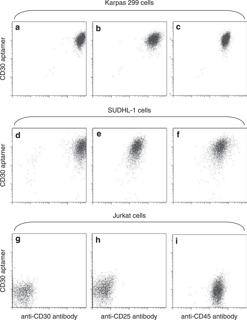
Figure 6.
Multicolor staining of cells by combined exposure to the CD30 aptamer and antibodies. Cultured Karpas 299 cells (a–c), SUDHL-1 cells (d–f), and Jurkat cells (g–i) were incubated with mixed probes, including the Cy5-labeled CD30 aptamer, PE-labeled anti-CD25 antibody, FITC-labeled anti-CD30 antibody, and AmCyan-labeled anti-CD45 antibody. Resultant cell staining was examined by multicolor flow cytometry analysis.

