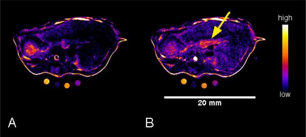
Fig. 4.
T1-weighted MR images of the mouse abdomen showing the axial view of the pancreas (duodenal side). Pre-GdDOTA-biPYREN (A), and 10 min post i.v. injection of the CA (B). Images were collected using a FSEMS sequence with the following parameters: TR = 89.03 ms; effective echo time (TE) = 11.21 ms; FOV 30×30 mm2, data matrix = 256×256, averaging = 3, slice = 1 mm, number of slices = 6, gap = 0; ETL=1, kzero = 1.

