
Figure 4. .
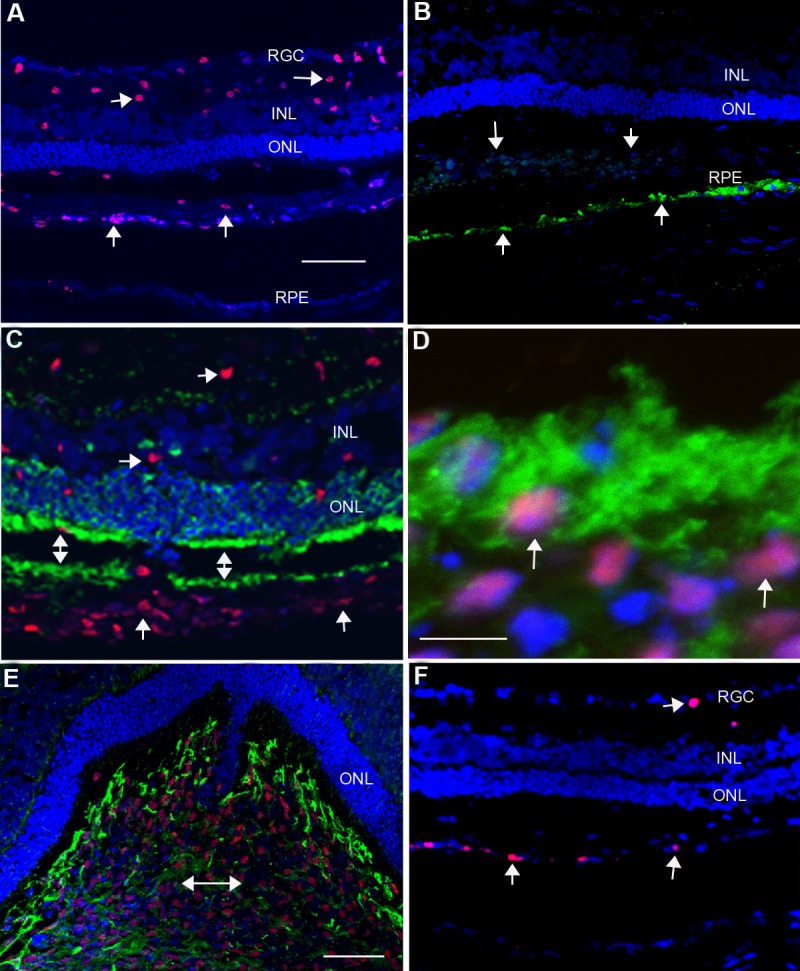
NSCs had two distinct locations: one formed a layer in the subretinal space (up pointing arrows in [A]); another one dispersed in the inner retina (right pointing arrows in [A]). There was robust photoreceptor preservation even at P180 (A). (B) Retinal section next to (A) stained with antibody RPE65 showed host RPE cells were positively stained (up pointing arrows in [B]), while donor cells were negative for RPE 65 (down pointing arrows in [B]). (C) Retinal section was double stained with human nuclear marker (red) and recoverin (green), there was no colocalization. (D) High power image showing donor cells (purple) were surrounded by recoverin positive materials (green). (E) Retinal section was double stained with human nuclear marker (red) and human nestin (green), it showed that donor cells expressed nestin. (F) Retinal section was stained with human specific antibody to Ki67, it showed a few positive stains in the subretinal space (up pointing arrows) and inner retina (right pointing arrow). Scale bars = 50 μm for (A) and (E), and 10 μm for (D).