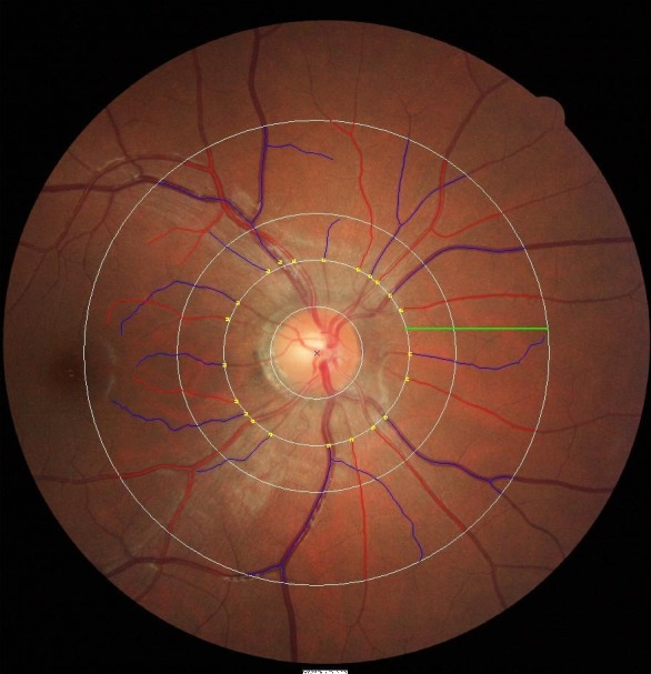
Figure. .

Retinal vessel measurements are performed 0.5- to 2.0-disc diameters away from the optic disc margin (zone C) by SIVA software, shown as a green line in the Figure. Estimates of central retinal arteriolar equivalent and central retinal venular equivalent, which represented the average diameters of retinal arterioles and retinal venules, were calculated by the revised Knudtson-Parr-Hubbard formula. Arterioles are marked in red and venules are marked in blue.
