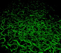
Movie.

3D SHG view of the DM of the GK cornea No. 7 in false colors (200 × 200 × 9 μm3). Strong SHG signals are emitted by fibrillar collagen abnormalities, which allows their spatial characterization. Note that the fibrils in the foreground appear larger than in the background because of the 3D viewer.
