
Figure 6. .
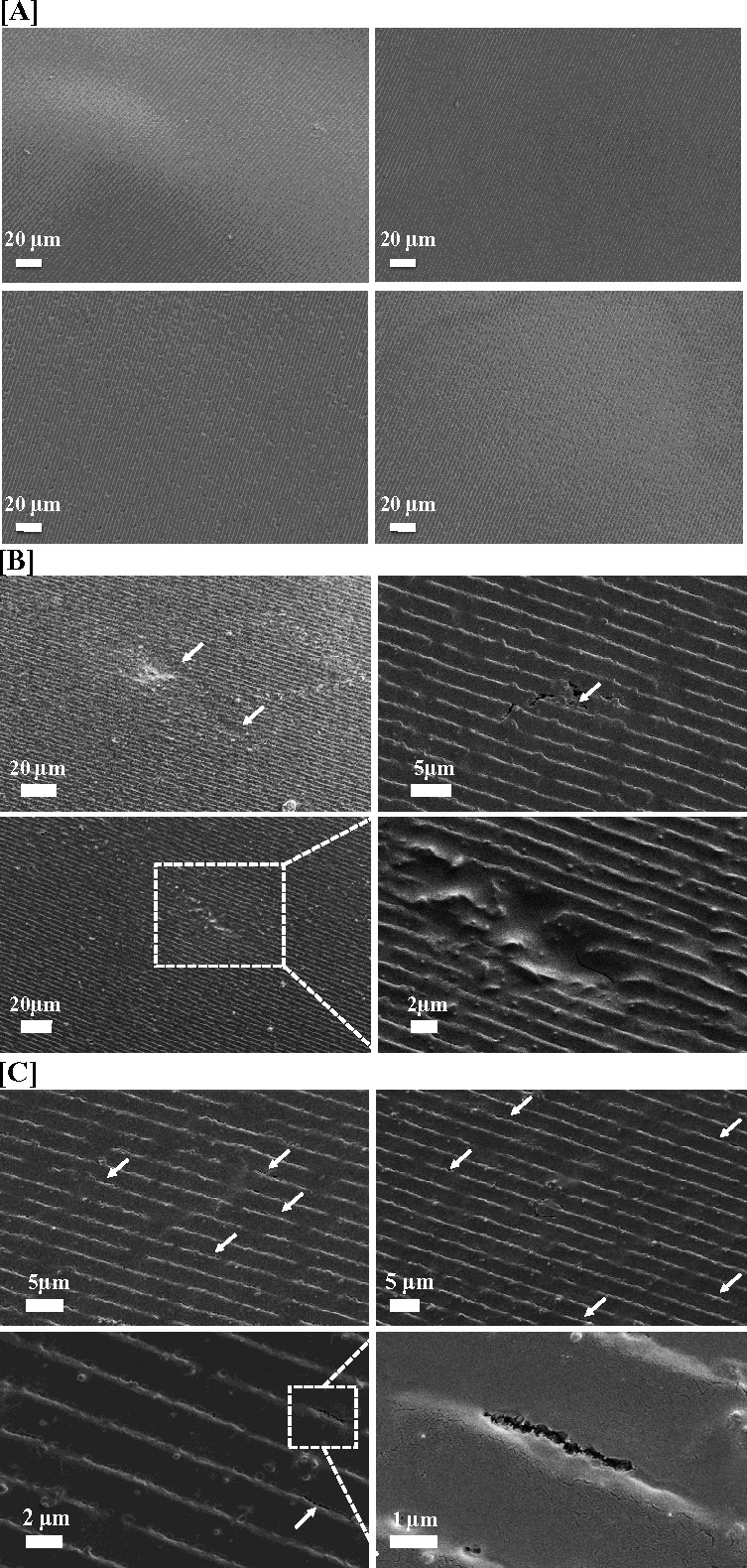
Scanning electron micrographs of the silk films after degradation with hCF for 8 weeks. (A) Control samples incubated in media with no cells, (B, C) samples incubated with hCF showing (B) degradation pits (arrows) and (C) degradation holes and cracks (arrows). One of the cracks was zoomed in the right bottom corner.