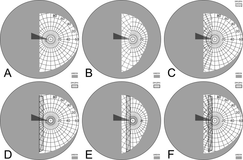
Figure 7. .
Building a percept diagram with confusion and diplopia. (A) The OD view, when the patient is gazing 20° into a 20Δ OS-only sector prism, is unaffected by the prism. (B) OS view. The view shifted through the prism brings 11° of the grid from the blind hemifield into view, while losing view of an 11° section at the apical scotoma. (C) Combined percept. There is visual confusion in the 20° section where OS sees through the prism and OD does not. Although OS views 20° of field through the prism, only the leftmost 11° provide expansion. The remaining 9° seen by OS at the prism apex side are visible to OD, but are seen in a different apparent direction, and thus diplopic. At the prism apex OD sees the 11° of field lost to OS by the apical scotoma. (The OD optic nerve head is outlined for clarity, but not likely perceived.) The OD-only view in (D) shades the portion of the OD field behind the OS prism that is seen diplopically, and the OS-only view in (E) shades the corresponding portion of the OS field. The field of view covered by the outlined areas in (D) and (E) is identical, but separated in the patient's view by the 11° prism power. In the final diagram (F), the outline for the diplopic region of only the prism view is shown, as that is the area in which diplopia is present without adding any expansion benefit, while the remaining areas of visual confusion are a necessary consequence of true field expansion. Central diplopia and confusion are annoying and confusing and, thus, poorly tolerated.

