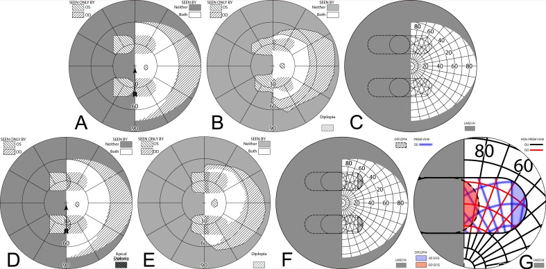
Figure 11. .
(A–C) Forward gaze with unilateral 57Δ EP Prism glasses. (D–F) The corresponding diagrams with 40Δ prisms. (A) Simulated Goldmann visual field. The left eye sees regions shifted from 30° left, while the right eye maintains a view of the regions blocked by the left eye's prisms' apical scotomas. Since the monocularly-viewed regions do not overlap, there is confusion but no diplopia, and there is a small gap between the expansion area and the normal seeing hemifield. (B) Actual composite field diagram from a patient. Imprecision in measurements or different fitting parameters result in a small region of apparent diplopia. (C) The corresponding percept diagram shows that there is visual confusion in the peripheral prism regions (where it is reasonably tolerated). The patient does not intentionally gaze into the prisms, but activity or objects seen there can alert the user and cause a gaze shift into the blind hemifield, possibly adjusting head direction slightly to view the area of interest centrally and not through the prisms. Dashed outlines indicate the source of the prism views. For simplicity and clarity, we plot rounded rectangles as the projected shape of the prisms. In actuality, they would appear slightly trapezoidal, with slightly curved horizontal edges. (D–F) With the lower-power prisms, the diplopic area, where the prism view includes portions of the normal seeing hemifield, is larger, since the prism power is less than the angular size of the visible portion of the prisms. Diplopia crosshatching in the clinical perimetry is based on the overlap found in monocular OS and OD diagrams, but was not actually reported by the subjects, which we take as another indication of its inconsequence peripherally. Diplopia in the percept diagram is outlined and lightly shaded in prism view locations where it offers no expansion benefit. (G) Annotated detail of the upper prism area of (F), with color coding to identify the contribution of each eye to the visual confusion and diplopia. The area behind the prism shaded in red (and its red grid lines) is seen directly by OD, while the same area (blue shading and blurred blue grid lines) is seen by OS shifted to a different location by the prism (diplopia).

