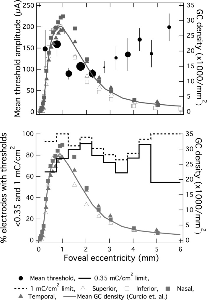
Figure 5. .
The effect of local ganglion cell density on stimulus threshold. (top) Mean threshold current amplitude (black circles) and ganglion cell density (grey connected symbols represent each retinal quadrant from Curcio and Allen's whole-mount study, solid grey line is the mean of these four quadrants) plotted against foveal eccentricity. Only electrodes in contact with the retina are included (in contrast with data plotted in Fig. 4), and electrodes are pooled across all subjects. Each solid black circle is representative of a 0.5 mm foveocentric bin with increasing diameter representative of electrode count for that bin. Vertical bars represent standard error. Mean threshold (black circles of varying diameter) and anatomical data correlated well (P = 0.03). (bottom) Percentage of electrodes with thresholds below 0.35 (solid line) and 1 mC/cm2 (dashed line) plotted against foveal eccentricity (0.5-mm bins up to 4 mm from fovea centralis; 1-mm bins at eccentricities greater than 4 mm to ensure minimum sample size was 10 electrodes/bin). Bin count ranged from 11 to 63 electrodes.

