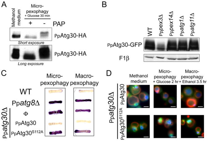
Figure 3. PpAtg30 Phosphorylation.
(A) PpAtg30 is a phosphoprotein. Ppatg30Δ cells expressing PpAtg30-HA (SJCF590) were grown 6 hr in methanol and micropexophagy was induced for 30 min. Three OD of cells were incubated for 20 min with and without PAP at 30° C.
(B) PpAtg30 phosphorylation depends on PpPex3. PpAtg30 phosphorylation was analyzed by immunoblotting of cell lysates expressing PpAtg30-GFP: wild-type (SJCF600), Pppex3Δ (SJCF330), Pppex14Δ (SJCF366), Ppatg1 D (SJCF473), and Ppatg11Δ (SJCF420).
(C) S112A mutation in PpAtg30 blocks pexophagy. Strains used for the AOX test are: wild-type (PPY12), Ppatg8Δ (SJCF257), Ppatg30Δ (SJCF44), Ppatg30Δ expressing PpAtg30-GFP (SJCF632), and Ppatg30Δ expressing PpAtg30S112A-GFP (SJCF736).
(D) The mutation S112A does not affect PpAtg30 localization. Ppatg30Δ cells coexpressing PpAtg30-GFP or PpAtg30S112A-GFP and BFP-SKL (SJCF385 andSJCF757) were analyzed by fluorescence microscopy.

