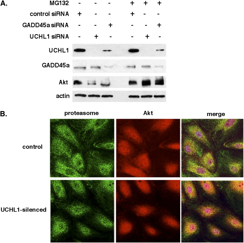
Figure 7.
Role of ubiquitin carboxyl terminal hydrolase 1 (UCHL1) and proteasomal degradation in Akt regulation by growth arrest and DNA damage–inducible α (GADD45a). (A) Western blots of endothelial cell (EC) transfected with silencing RNA (siRNA) specific for GADD45a or UCHL1 (100 nM, 3 d) reveal comparable decreases in total Akt levels compared with controls that were similarly reversed by treatment with the proteasomal inhibitor MG132 (10 μM, 1 h). Notably, silencing of GADD45a was also associated with a significant decrease in UCHL1 protein levels. (B) Immunofluorescence of EC reveals a minimal association of Akt with proteasome (20S) under control conditions (merge, top panel). Conversely, in EC transfected with siRNA specific for UCHL1 (100 nM, 3 d) there is a marked colocalization of Akt with proteasomes (indicated by yellow in the merge image, lower panel) consistent with Akt proteasomal degradation in UCHL1-silenced cells.

