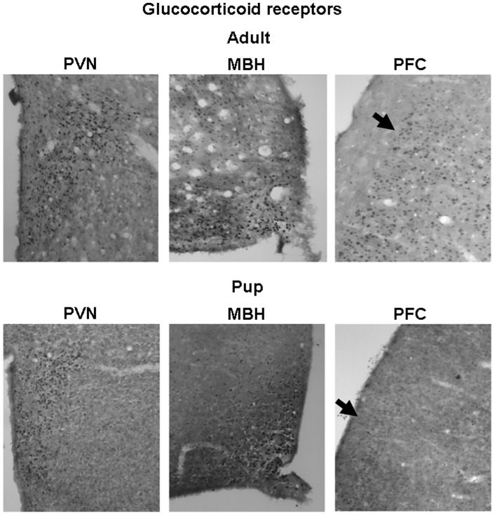
Figure 4. Representative pictures of glucocorticoid receptor immunohistochemistry on adult (approx. 3-month-old) and pup (10-day-old) rat brains.
Selected area of the hypothalamus (PVN: nucleus paraventricularis hypothalami, MBH: mediobasal hypothalamus) and prefrontal cortex (PFC) are presented. Arrows represent different distribution of the GR immunoreactivity in adult and pup PFC regions.

