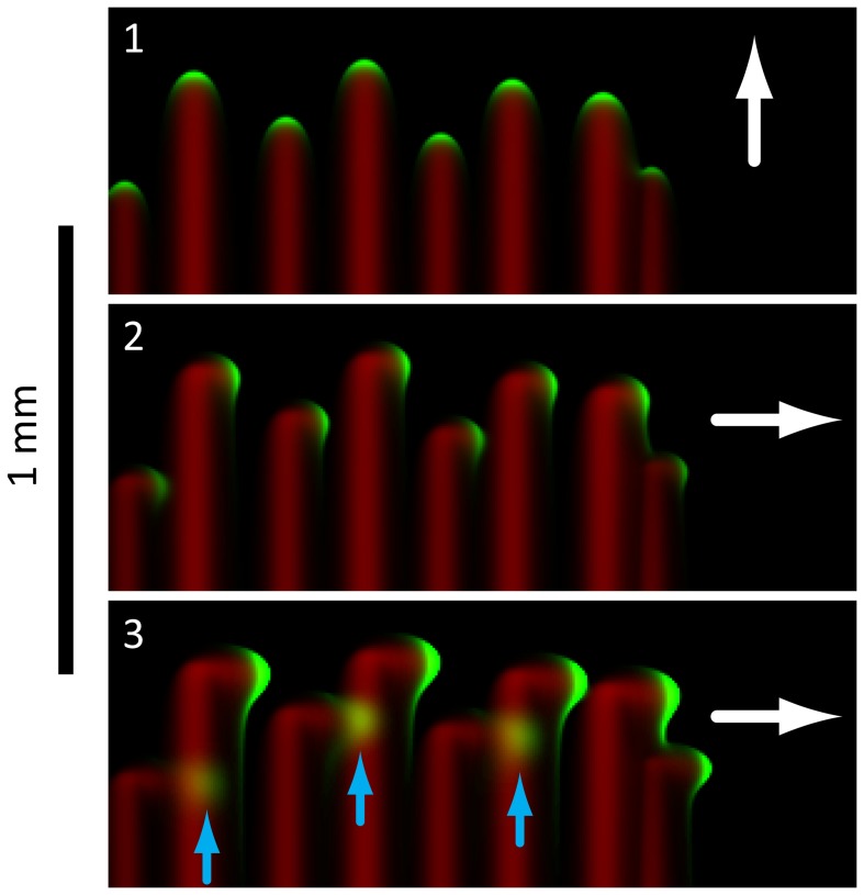
Figure 4. Biophysical model reproduces key features of group motility during finger merging.
In these simulations, the direction of light bias (white arrows) is rotated 90 degrees between frames 1 and 2. The cells (green) leave an EPS trail (red), and when a finger intersects the EPS trail left by a neighboring finger, the group of cells speeds up and spreads out (blue arrows, frame 3). We observed the same qualitative changes in finger merging experiments (Fig. 2).

