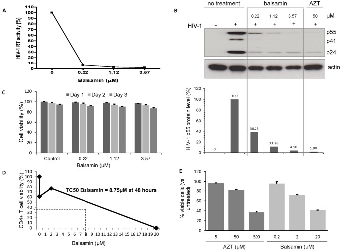
Figure 3. Balsamin potently inhibits HIV-1 replication in primary CD4+ T cells.
A. Primary CD4+ T cells were infected with HIV-1 at a moi of 0.1, in the absence or presence of indicated amounts of balsamin. Eight hours post-infection, cells were washed with PBS, and incubated further with relevant amounts of balsamin. Three days post-infection, cell free supernatant was harvested for assessing viral replication by RT assay. The values obtained in the absence of balsamin were arbitrarily set as 100%. B. In parallel, cell lysates were collected and were immunoblotted for HIV-1 p24 (B, upper panel). Actin served as a loading control, and AZT was used as a positive control for the HIV-1 inhibition. The intracellular level of p55 was quantified and plotted (B, lower panel). The sections of this figure are representative of three donors. C. In parallel of this assay, putative cytotoxic effect of these different concentration of balsamin on primary CD4+ T cells were monitored by determining the percentage of viable cells using Trypan blue exclusion. Data are representative of three donors and experiments performed in duplicate (±SD). D. Primary CD4+ T cells were incubated for 48 hours with balsamin at 0.2 µM, 2 µM and 20 µM. At 48 hours cells were harvested for determination of cell viability by trypan blue assay and cell counting. One representative experiment out of two is shown. E. Primary CD4+ T cells were treated as above in parallel with AZT and stained for Annexin-V and 7-AAD. Viable cells (Annexin-V−/7-AAD−) were measured by FACS and plotted on a bar graph +/− SD (n = 2 in duplicate).

