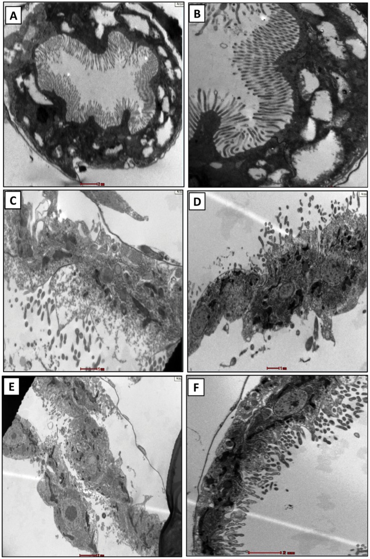
Figure 11. Transmission electron microscopy of the alimentary canal.
Alimentary canal of untreated daphnids shows (A) uniform gut lining with intact microvilli and basal membrane; (B) a closure view of the gut lining showing healthy microvilli and basal membrane. The treated samples show (C) disrupted cellular features with (D) disintegrated micrivilli and basal membrane. Certain samples also showed (E) complete destruction of microvilli with few loosely attached cells and (F) abnormal cellular interior. BM: basal membrane; MV: microvilli. The gut lining of at least five different animals were observed to draw a conclusion (n = 5).

