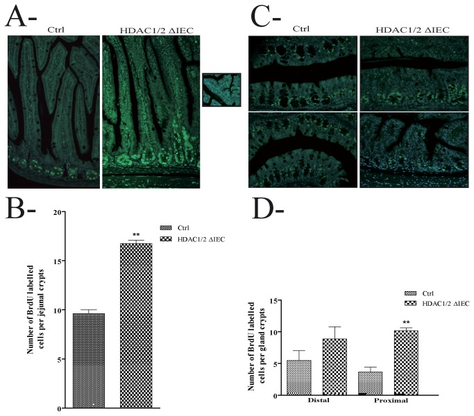
Figure 3. Conditional intestinal epithelial HDAC1/2 loss leads to increased proliferation.
2 h after BrdU intraperitoneal injection, four-month-old jejunal (A) or colonic (B) tissue sections from control (Ctrl) or conditional intestinal epithelial HDAC1/2 (HDAC1/2ΔIEC) mice were revealed with an antibody against BrdU. The insert in A shows the absence of BrdU-labelled cells in branched villi. The average number of BrdU-labelled cells per jejunal (C) or proximal and distal colonic (D) crypts was measured (n=3; 20 to 30 crypts each). Results represent the mean ± SEM (**p≤0.01). Magnification: 20 X.

