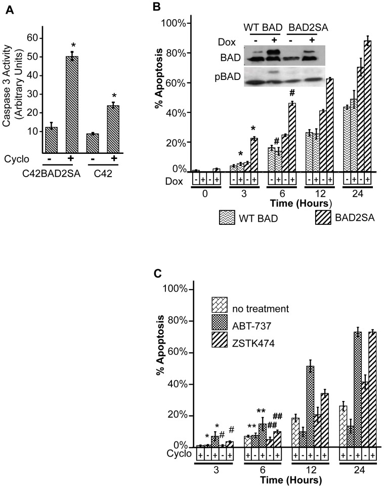
Figure 4. Phosphorylation-deficient BAD and a BAD mimetic sensitizes prostate cancer cells to apoptosis by protein synthesis inhibitors.
A) Caspase 3 activation in C42Luc and C42LucBAD2SA cells that expressed a phosphorylation-deficient BAD mutant. Cells were treated with 100 µg/mL cycloheximide for 6 hours before assay. *, p<0.01. B) Analysis of apoptosis by time-lapse microscopy. C42Luc cells were transiently transfected with a doxycycline-inducible HA-BAD2SA or wild-type HA-BAD, and treated with doxycycline (Dox) 48 hours after transfection. After 18 h of Dox treatment, cells were treated with 100 µg/mL cycloheximide, and the cumulative percentage of apoptotic cells was determined by time lapse microscopy. At least 100 cells were counted for each treatment. Error bars show standard deviations from the average of four randomly chosen fields. *, p<0.01 #, p<0.03. Inset shows Western blot analysis of phosphorylated BAD (pBAD112) and total BAD expression in C42Luc cells collected 18 hours after Dox treatment. Lower bands correspond to endogenous protein, while the upper bands correspond to inducibly expressed HA-BAD constructs. C) C42Luc cells were treated with 100 µg/ml cycloheximide in combination with 10 µM ABT-737, a BH3 mimetic based on the BH domain of BAD or ZSTK474. Apoptosis was analyzed by time-lapse microscopy. Cumulative cell death at indicated time points is shown. At least 100 cells were counted for each treatment. Error bars show standard deviations from the average of four randomly chosen fields. *, p<0.01 **, p<0.05 #, p<0.001 ##, p<0.002.

