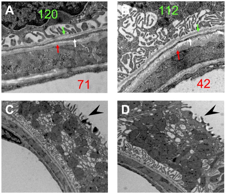
Figure 3. EM observations on the nonA-nonB intercalated cells at contact sites.
(A) and (B) are the high magnification images of the framed areas in Figure 2 (nephron Nr. 120 and Nr. 112). The fibroblast extensions (white arrows) interpositioned between the basement membranes of the Af-Arts (red arrows) and the tubules (green arrows). Note the membranous infolding structure on the base of the IC. (C) and (D) are other two examples of the intercalated cells at the contact sites. Note the microprojections on the luminal surface (arrow heads) and the numerous mitochondria and vesicles that are not restricted to the apical area of the cell. (A), (B) ×15,000; (C), (D) ×10,000.

