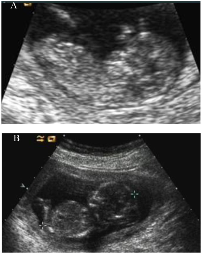
Figure 3. Ultrasound image of the normal and abnormal fetuses.
A: The ultrasound image of PGD fetus at the 12 weeks’ gestation showing intact skull and did not have an enlarged abdomen. B: The ultrasound image of the third pregnancy of this couple at the 14 weeks’ gestation showing occipital encephalocele and enlarged abdomen.

