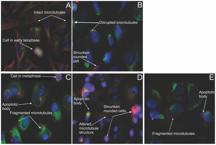
Figure 11. Immunofluorescent determination of microtubule dynamics in HeLa cells.
Double immunoflourecence was conducted to determine the compounds' effect after 24 h on microtubule dynamics within HeLa cells. Tyrosinated (dynamic) microtubules are visualized as red, whereas the detyrosinated (stable or stabilized) microtubules are stained in green. The vehicle-treated cells (A) demonstrated an intact dynamic microtubule structure. Both the colchicine positive control (B), as well as the ESE-16-treated cells (C) showed complete microtubule depolymerisation with few detyrosinated microtubule fragments remaining. ESE-15-one-treated cells (D) demonstrated altered microtubule morphology. EMBS-exposed cells (E) revealed fragmented microtubules. All treated cell samples demonstrated a decrease in cell density and rounded cells (X63 oil objective).

