
Figure 9. Sequence alignment and annotated tertiary motifs in Mg2+ riboswitches.
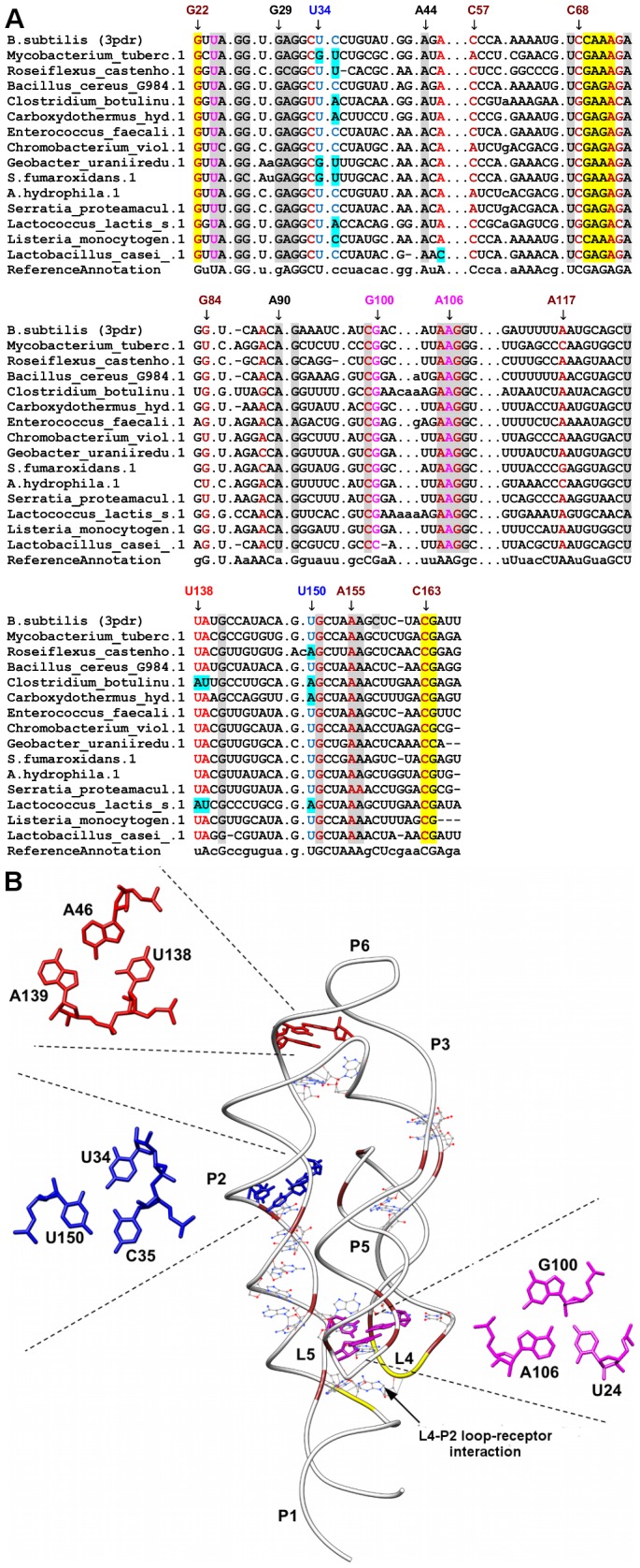
(A) Sequence alignment of magnesium riboswitches where gray shaded columns indicate the highly conserved nucleotide positions. Other color shaded columns indicate regions involved in tertiary interactions (brown: A-minor and yellow: loop-receptor interaction). For clarity, not all tertiary motif positions are numbered. Base-triple positions that are not conserved are in cyan shaded columns. (B) Tertiary structure of B. subtilis Mg2+ [26] riboswitch (PDB ID = 3pdr). The ligand is represented as spheres and the highly conserved nucleotides are in wire representation. Magnifications of the triples using numbering for 3pdr are presented in red (A46-U138-A139), blue (U34-C35-U150) and magenta (U24-G100-A106).