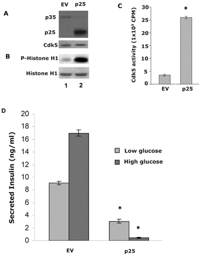
Figure 3. Min6 cells infected with p25, display high levels of Cdk5 activity and inhibits insulin secretion.

A Expressions of infected p25 (panel 1, Lane 2) and endogenous p35 (panel 1, lanes 1 and 2) and Cdk5 (panel 2 lanes 1 and 2). B Autoradiograph of phosphorylated histone H1 in infected Min6 cells. The same cell lysates were used for the kinase assay using Cdk5 immunoprecipitates. C Quantification of Cdk5 activities in infected Min6 cells. Data represent mean ± SE of three experiments. D Insulin secretion was extremely inhibited when p25 were overexpressed in Min6 cells. Insulin released from p25 infected cells was dramatically inhibited under basal and stimulated conditions, more than 95% in the latter case, (* P<0.01). Data represent mean ± standard error of three experiments.
