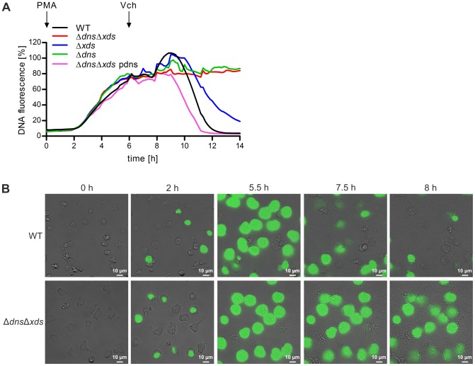
Figure 3. The two extracellular nucleases of V. cholerae are able to degrade NETs.
A. DNA release of neutrophils was stimulated with PMA (indicated by the arrow “PMA”) and followed by incubation with the respective V. cholerae strain (MOI 40, time point of addition is indicated by the arrow “Vch”). Staining of DNA by the cell impermeant fluorescent DNA dye Sytox green was measured in 10 min intervals. Values are presented as percentage of DNA fluorescence compared with the Triton ×100 lysis control (100%) indicating NET formation, respectively. Shown are medians of at least six measurements out of three independent donors. B. Human neutrophils were stimulated with V. cholerae WT or ΔdnsΔxds mutant (MOI 4) in presence of the cell impermeant fluorescent DNA dye Sytox green and monitored by live cell imaging. Shown are images of the indicated time points. The complete movies are available as movie S1 (V. cholerae WT) and S2 (ΔdnsΔxds mutant) in the supporting information.

