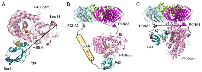
Figure 7. Models for binding between PdX and P450cam in PUPPET.

(A) A docking model of P450cam and PdX. The docking program GRAMM-X [42] was used to generate the model from crystal structures of P450cam (PDB: 1DZ4) and PdX (PDB: 1XLP) according to a previous report [40]. (B, C) Spatial arrangement of P450cam and the PCNA ring when the PdX-binding site of P450cam faces (B) in the same direction as or (C) in a perpendicular direction to the PCNA ring. The distance between the C-termini of PCNA2 and PCNA3 was estimated from the crystal structure of the PCNA heterotrimer (PDB: 2NTI).
