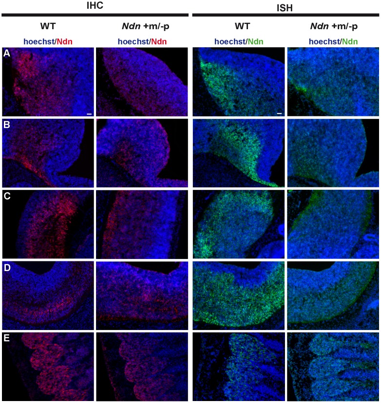
Figure 2. Ndn expression in Ndn+m/−p E12.5 embryos.
Expression of Ndn in the nervous system of WT and Ndn+m/−p embryos at E12.5 revealed by IHC or ISH on frozen sections using a Necdin specific antibody (Ndn,red) or Ndn RNA probe (green). Tissue sections are visualized using a Hoechst labeling (blue). Expression is detected in both genotypes, at the protein and transcript levels, in the preoptic area (A), supraoptic area (B), thalamus (C), pons (D) and in the dorsal root ganglia (E). Note that the level of expression is weaker and more restricted in Ndn+m/−p embryos. Other structures, like the tegmentum, the subthalamus and the spinal cord also express the Ndn maternal allele. Scale bar: 50 µm.

