
Figure 5. SPCLIP1 and TEP1 interact after challenge with E. coli bioparticles.
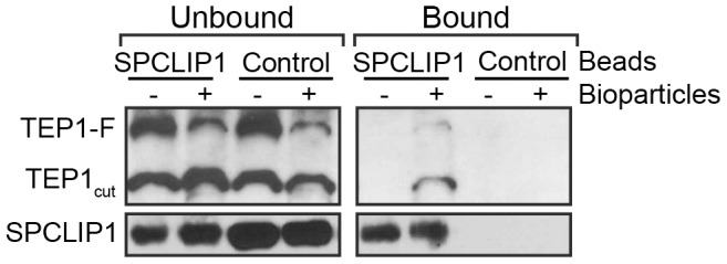
IP beads containing SPCLIP1 antibody or control beads were used to capture proteins from hemolymph 15 min after injection with E. coli bioparticles (+) or PBS (−). The beads were separated and samples of the unbound and bound fractions were analyzed by western blot under reducing and non-reducing conditions for TEP1 and SPCLIP1, respectively. Images are representative of two independent biological replicates.