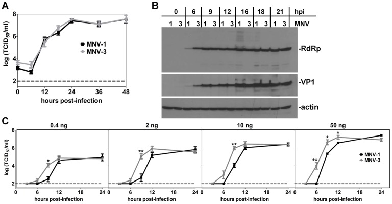
Figure 6. MNV-3 initiates replication faster than MNV-1.
A) RAW 264.7 cells were infected with MNV-1 or MNV-3 at MOI 5. Supernatant was collected from two independent wells at the indicated hours post-infection (hpi) and virus titers determined using TCID50 assay. The entire experiment was repeated three times and data from all experiments are averaged. The limit of detection of the assay is indicated by a dashed line. B) Infected cells from the same cultures used for panel A were lysed and viral proteins were detected by western blotting using the indicated antibodies. These data are representative of duplicate samples tested from each of three independent experiments. C) 1.5×10⁵ HEK-293T cells were transfected with 0.4, 2, 10 or 50 ng of purified MNV-1 or MNV-3 genomic RNA. The virus titers at the indicated hpi were determined using TCID50 assay. Data for all replicates are averaged. Data for MNV-1 and MNV-3 at each time point were compared for statistical purposes.

