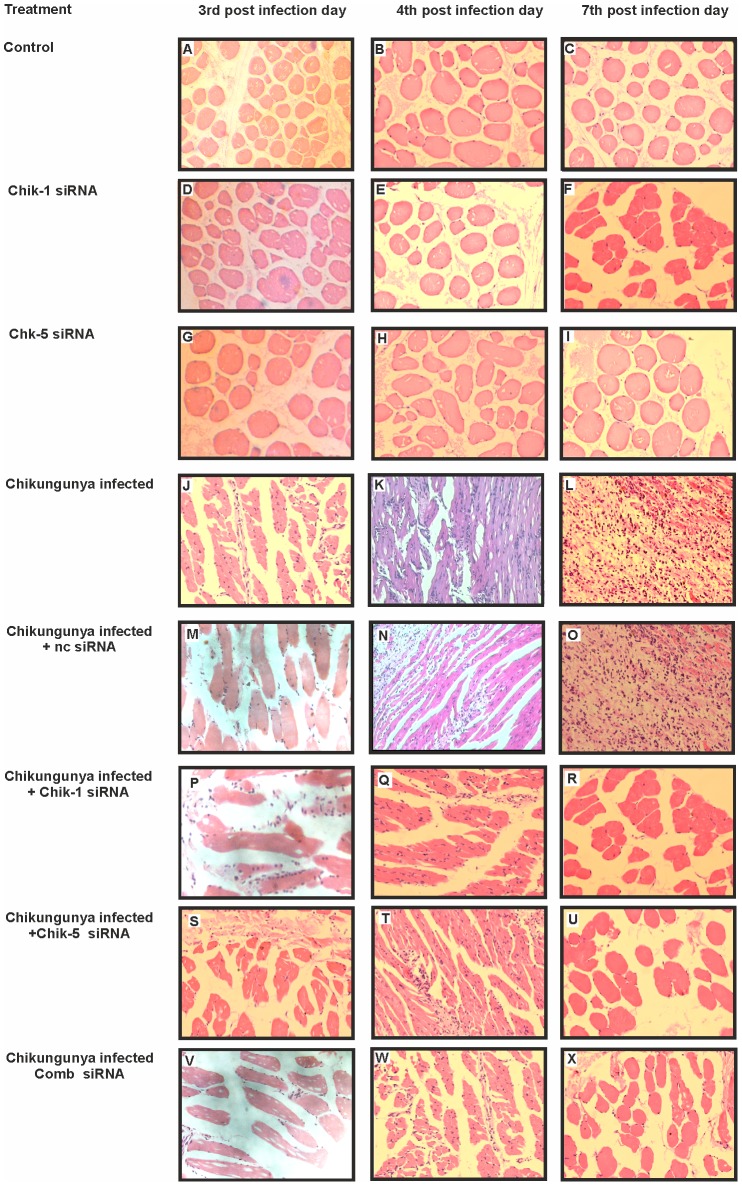
Figure 10. Histopathological changes in mouse muscle tissues after chikungunya infection and siRNA treatment.
C57BL/6 mice were infected with CHIKV i.v. (1×10⁶ PFU CHKV; 100 µl of 107pfu/ml). Hematoxylin/eosin-stained tissue sections were screened to investigate the pathological effects of siRNA treatment. PBS injected mice showed normal cellular organization (A, B and C). No significant cellular changes were observed in E2 siRNA treated mice (D, E and F) and ns1 siRNA treated mice (G, H and I). At 3 days p.i. mild inflammation and mild monocyte/macrophage infiltrates were observed (J, M, P, S and V). At 4 days p.i. and 7 days p.i. CHIKV infected and ncsiRNA treated muscles showed pronounced monocyte/macrophage infiltrates, necrosis and edema (K, L, N and O), whereas siRNA treated CHIKV infected mice muscle tissues showed the regeneration after treatment (Q, R, T, U, W and X) (Magnification ×400).

