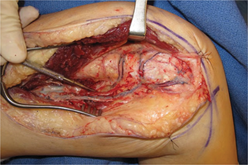
Fig. 1.

Medial view of femur after elevation of the vastus medialis. Longitudinal coursing DGA vessel is seen running from superficial femoral artery on left to MFC on right. Note the network of vessels on the medial femur, including the longitudinal vessel directed to the condyle and the transverse branch running toward the patellofemoral joint. This exposure is much more extensive than that required for the scaphoid reconstruction, but it is provided to demonstrate the vascular anatomy of the DGA system.
