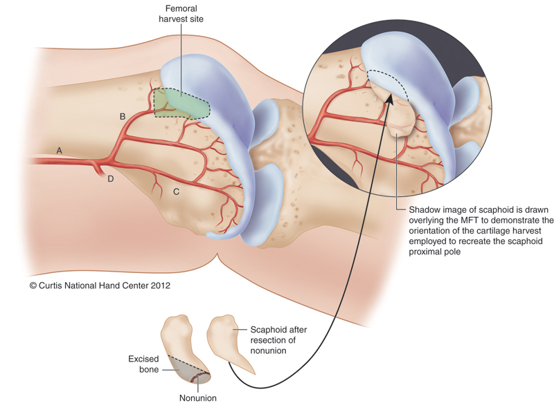
Fig. 3.

Representation of MFT and the planned portion of reconstructed proximal scaphoid. Portion of MFT harvested to provide vascularized osteocartilaginous reconstruction of the proximal scaphoid. A, descending geniculate artery; B, transverse branch; C, longitudinal branch; D, superiomedial geniculate artery. (From Bürger HK, Windhofer C, Gaggl AJ, Higgins JP: Vascularized medial femoral trochlea osteocartilaginous flap reconstruction of proximal pole nonunions. J Hand Surg Am 2013 April; 38(4):690-700).
