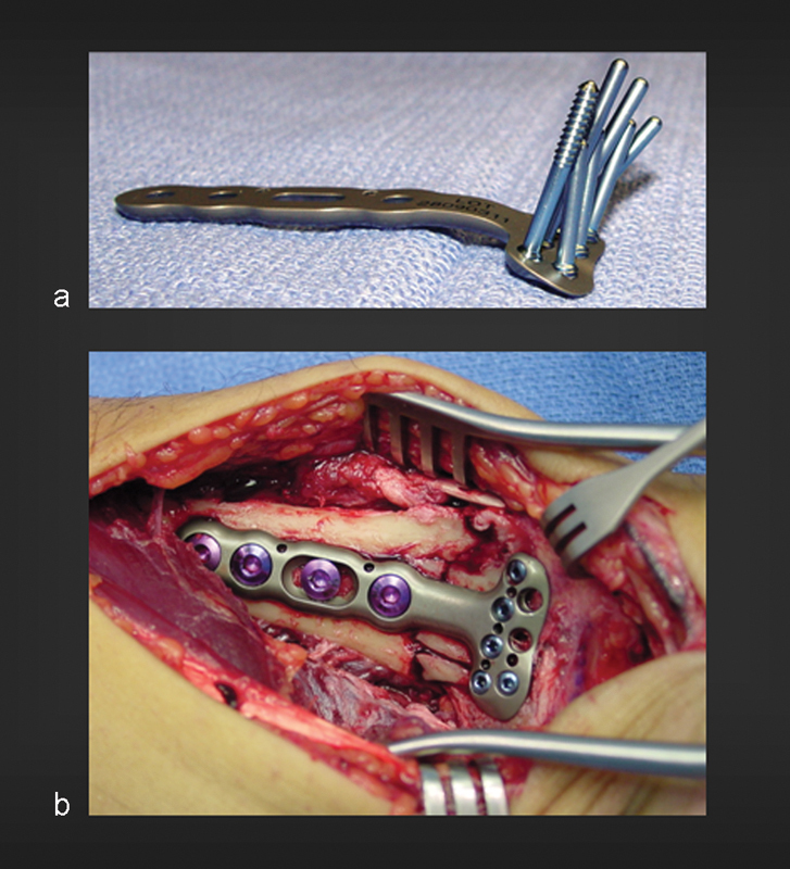
Fig. 1.

(a) DVR plate, showing the subchondral support pegs that buttress both the radial and intermediate columns of the affected articular surface. Notice the fan-shaped distribution of the pegs to conform to the anatomy of the subchondral plate. (b) DVR plate in situ in a patient with intra-articular fracture of the distal radius, exposed through a volar approach. Notice the position of the plate just proximal to the watershed line.
