
Fig. 2.
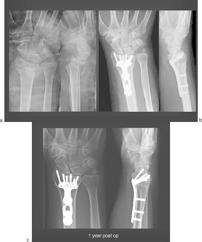
(a) X-ray images of a C3.1 intra-articular fracture with depressed lunate facet and dorsal displacement. Notice additional fracture at the base of the ulnar styloid. (b) Post-operative X-ray image showing anatomical reduction of the joint surface and restoration of length following volar fixed-angle plate fixation. (c) X-ray image at 1 year follow-up showing well-maintained joint surface and asymptomatic nonunion of the ulnar styloid.