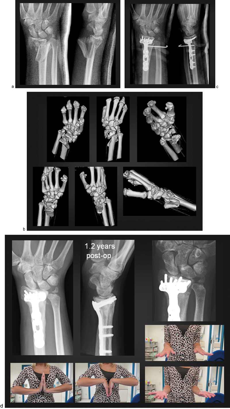
Fig. 3.

(a) X-ray images showing a high-energy intra-articular fracture of the distal radius, C3.3/Fernandez 5. (b) Three-dimensional (3D) computed tomography (CT) reconstruction revealing the extent of both the articular and metaphyseal comminution of the distal radius. Both the radiocarpal and the sigmoid notch articular surfaces are involved. Notice severe displacement of the ulnar styloid fracture. (c) Following anatomical reduction of the radius and the application of the DVR plate, the DRUJ was unstable and therefore was pinned to the radius (1.6 mm K-wire). The pin was left in place for a period of 3.5 weeks. (d) X-ray images at 1.2 years following the operation, revealing a congruent radiocarpal joint and a slight widening of the scapholunate interval. The DRUJ was noted to be well aligned, with persistent fibrous union of the ulnar styloid. Clinical photographs show almost symmetrical flexion, extension, pronation, and supination of the affected right wrist.
