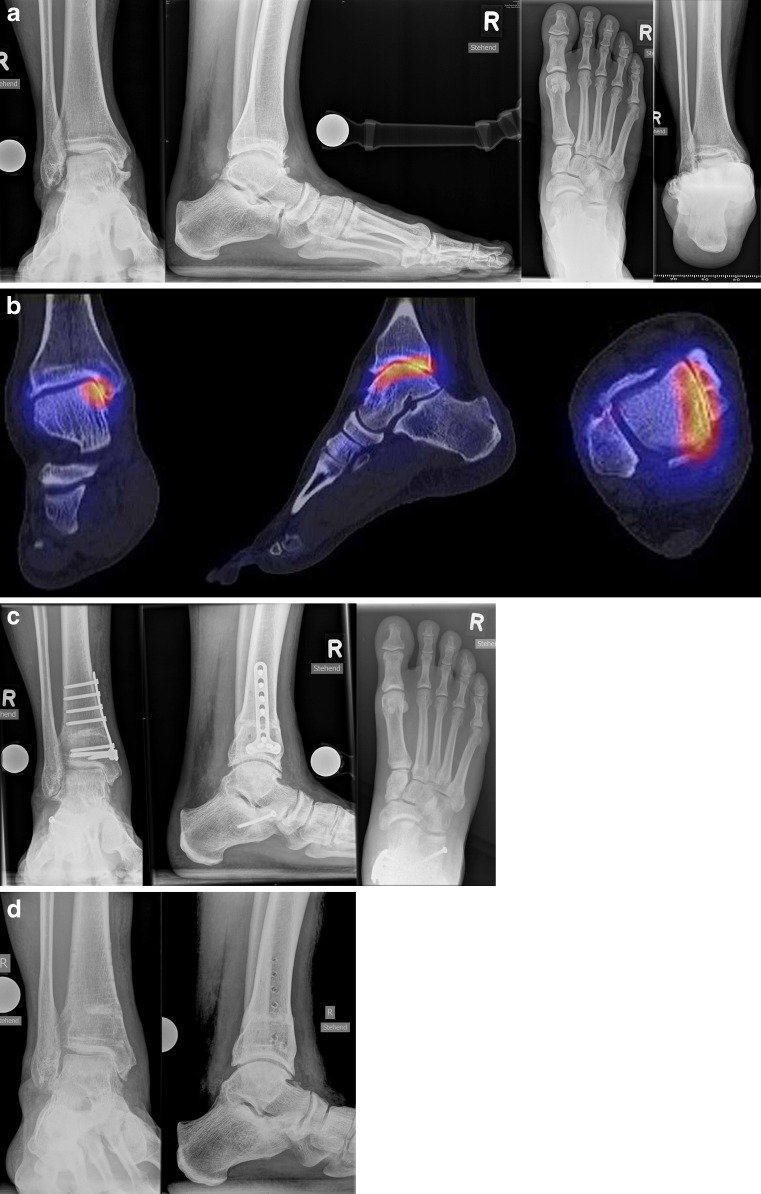
Fig. 5.
Medial opening wedge osteotomy. a Pre-operative weight-bearing radiographs show varus tilting of the talus within the mortise. However, the Saltzman view shows the valgus heel position, as the patient has peritalar instability with Z-shaped hindfoot deformity. b SPECT/CT shows biologically active degenerative changes of the medial tibiotalar joint. c Supramalleolar medial opening wedge osteotomy was performed to address the varus tilt of the talus and lateral lengthening calcaneal osteotomy to address the inframalleolar valgus deformity of the hindfoot. Post-operative weight-bearing radiographs show completed osseous healing at the site of osteotomies at the 1-year follow-up. d After hardware removal patient is pain-free with no restrictions of sports activities

