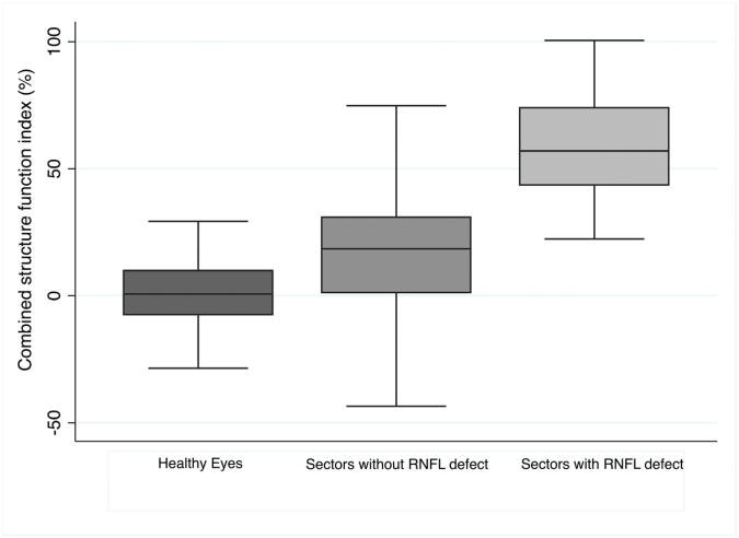
Figure 3.
Box plot showing the distribution of estimated retinal ganglion cell loss (estimated using the combined structure function index (CSFI)) in all sectors of healthy eyes compared to the combined structure function index in sectors with visible localized retinal nerve fiber layer (RNFL) defects and the combined structure function index in sectors of glaucomatous eyes without visible localized RNFL defects.

