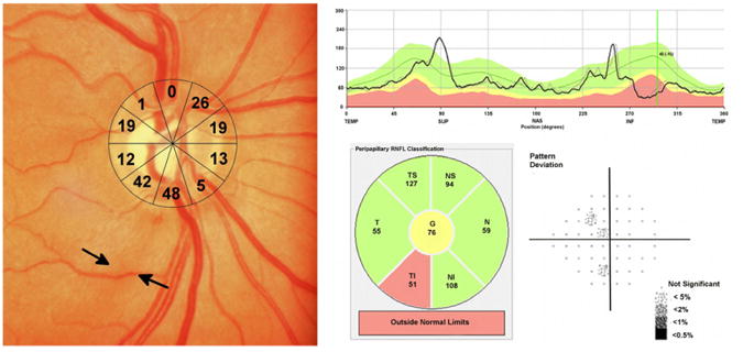
Figure 6.

Example of an eye of a 70 year-old subject with a localized retinal nerve fiber (RNFL) defect visible on optic disc photography.(Left) The RNFL defect corresponded to two sectors of the structure function map with an estimated retinal ganglion cell loss of 42 to 48%.(Left) The estimated retinal ganglion cell loss in the other sectors is also shown.(Left) Spectral domain optical coherence tomography revealed localized thinning of the RNFL in the inferior-temporal region.(Top right and Bottom center) Despite high estimated retinal ganglion cell losses, standard automated perimetry global indices were within statistically normal limits.(Bottom right)
