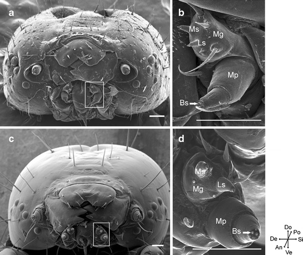
Fig. 3.

Scanning electron micrographs of fifth instar heads showing maxillae. a, c Anterior images of Bombyx mori (a) and Helicoverpa armigera (c) heads. b, d Enlarged views of a maxilla of B. mori (b) and H. armigera (d). White rectangles indicate the location of maxillae. Mg, maxillary galea; Ms, medial styloconic sensillum; Ls, lateral styloconic sensillum; Mp, maxillary palp; Bs, basiconic sensillum. Do, dorsal; Po, posterior; Si, sinistral; Ve, ventral; An, anterior; De, dextral. Scale bar: 100 μm
