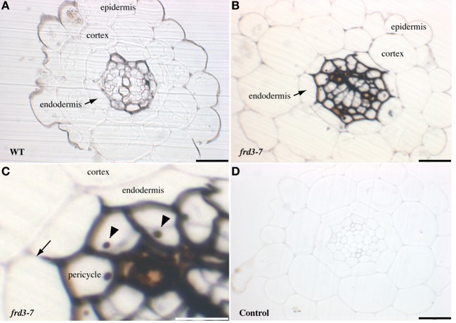
Figure 1.
Iron detection in Arabidopsis roots. Sections of 4-week old WT (A) or frd3-7 (B–D) mutant plants stained with Perls/DAB. Panel (C) corresponds to a magnified image of panel (B), both showing iron restriction within the boundary of the Casparian strip (arrow), nucleoli are indicated by arrowheads. (D), control section stained with DAB without previous Perls reaction. Scale bars: 50 μm (A,B,D) or 25 μm (C).

