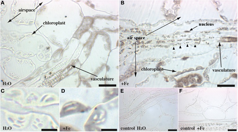
Figure 2.
Iron excess in Arabidopsis rosette leaves. Sections of 4-week old WT plants irrigated with either water (A,C,E) or Fe-EDDHA 2 mM during 48 h (B,D,F) were stained with either Perls/DAB (A–D) or DAB alone as a negative control (E,F). The panels (C,D) correspond to a magnified image of regions of panels (A,B), respectively. Arrowheads in panel (B) indicate Fe-rich structures in the vascular tissues. Scale bars: 20 μm (A,B,E,F) or 5 μm (C,D).

