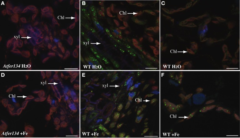
Figure 4.
Immunolocalization of Ferritin in Arabidopsis leaves. Sections of rosette leaves from 3 week-old WT (B,C,E,F) or triple mutant Atfer1,3,4 (A,D) plants irrigated with either water (A–C) or 2 mM Fe-EDDHA (D–F) were probed with an anti-ferritin antibody and revealed with a secondary anti-rabbit antibody coupled to the Alexa Fluor® 488 fluorophore. Sections were stained with DAPI to reveal cell nuclei. Ferritin localization appears in green, DAPI fluorescence in blue and chlorophyll auto-fluorescence in red. Scale bars: 10 μm.

