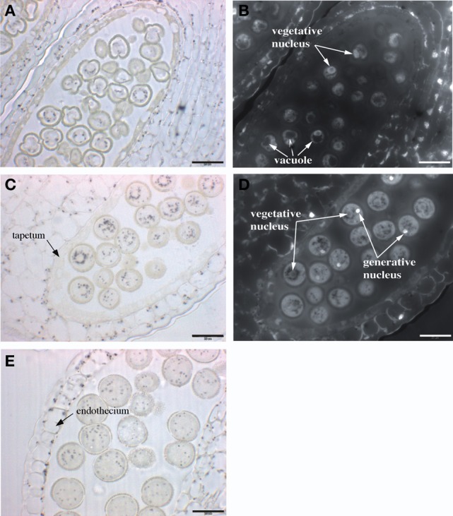
Figure 6.

Iron distribution during Arabidopsis pollen development. Sections of anthers at three different stages of pollen development were stained with Perls/DAB and DAPI. (A,B) mononuclear pollen grain, (C,D) binuclear pollen grain, (E) mature pollen grain. (A,C,E) Bright field images; (B,D) Epifluorescence images from slides (A,C), respectively, showing DAPI-stained vegetative and generative nuclei. Scale bars: 20 μm.
